Temperature screening thermography uses a variety of devices ranging from low-end handheld scanners up to medical-grade devices with the same detailed specifications required for evaluation of human and veterinary patients. High-end temperature screening systems use facial recognition software to calculate body temperature from the medial canthus of the eye, which is the most accurate superficial point for evaluating core body temperature.1 These systems measure temperatures from six feet away, allowing for safe, contactless use. Further, they "flag" elevated temperature; the person then undergoes a secondary screening protocol.
Medical thermography is more than 70 years old. Early devices were expensive, cumbersome, and not practical for widespread clinical application. In 1980, reports began validating thermal imaging as a tool for the evaluation of musculoskeletal conditions in horses.2,3 By 2001, multiple publications had established the technology as a practical adjunct to clinical and radiological examinations in assessment of equine lameness.4,5,6,7
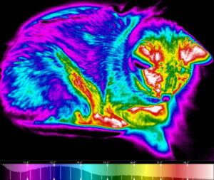
In veterinary medicine, thermal imaging has garnered increased interest over the last decade as veterinary-specific digital devices became available. Thermal images now give us new information that is as important as what we learn from vital signs we rely on. Traditional vital signs are important measures of the patient's physiological status and are part of a pattern of health or disease. Thermal images give us a new, additional vital sign—they are a visual presentation of body surface temperatures and are representative of temperature distributions in the tissues below.8 They give us physiological information and serve as a road map for further diagnostics.